
OEM Stress Test ECG Electrodes | Diaphoretic Cardiac Sensors | TOP-RANK
B2B contract manufacturing of stress test and diaphoretic ECG electrodes for treadmill ergometry and emergency triage. Features offset snap geometry, quick-wetting liquid gel (<100 Ohm at 10 Hz), aggressive hydrophobic acrylic adhesive (>6.5N/25mm), and ANSI/AAMI EC12 compliance.
Product Overview
Stress test and diaphoretic ECG electrodes operate as disposable diagnostic biopotential interfaces designed for high-motion, high-perspiration clinical protocols. These include Bruce protocol treadmill tests, cycle ergometry, nuclear myocardial perfusion imaging (MPI), pharmacological cardiac stress tests, and emergency department triage of diaphoretic acute coronary syndrome (ACS) patients. The assembly isolates biopotential voltages (0.5 mV to 2.0 mV) generated by myocardial depolarization while withstanding continuous patient vibration, chest wall expansion, and profuse eccrine perspiration.
Standard diagnostic monitoring electrodes frequently fail during cardiac stress protocols across three specific mechanisms:
- Motion-Induced ST-Segment Distortion from Centered-Snap Shear: Traditional electrodes locate the brass snap directly over the conductive gel core. During running (acceleration forces reaching 10 to 15 m/s²), heavy patient lead cables whip cyclically. This kinetic energy transfers down the snap into the sensing layer, deforming the Helmholtz electrical double layer (Vhc) at the skin-electrolyte boundary. The resulting potential fluctuation creates low-frequency motion artifacts (0.05 Hz to 1.0 Hz) and baseline wandering that mimics or obscures ST-segment depression (>0.1 mV), triggering false ischemia readings.
- Adhesive Plasticization and Sweat Gland Delamination: Under physical exertion, eccrine sweat glands produce water and sodium chloride at rates up to 2.0 liters/hour. Standard medical adhesives absorb this aqueous fluid, undergoing hydro-plasticization and losing shear resistance. As sweat wicks between the tape and the stratum corneum, the electrode border detaches, causing loss of signal midway through maximal exertion (Stage 4 or 5 of the Bruce protocol).
- Impedance Lag from Slow-Hydrating Hydrogels: Solid hydrogels rely on slow passive moisture transfer to wet the dry stratum corneum, requiring 5 to 10 minutes to reach low impedance. If stress testing begins immediately after application, the high initial interface impedance (Rct > 3,000Ω) forces the ECG preamplifier into baseline wandering and high-frequency noise pickup.
This electrode platform resolves these issues through an offset (eccentric) geometry that separates the snap terminal from the conductive well. The contact interface features an aqueous, high-chloride liquid gel impregnated into an open-cell reticulated polyurethane foam sponge, achieving full stratum corneum wet-out in under 15 seconds. The carrier uses a 1.0 mm high-density, closed-cell cross-linked polyethylene foam coated with a hydrophobic acrylic adhesive delivering dynamic peel adhesion exceeding 6.5N / 25mm. We contract-manufacture these sensors for stress test equipment brands, cardiology clinic distributors, and emergency medical consumable networks globally.
Technical & Engineering Description
The industrial manufacture of stress test and diaphoretic ECG electrodes takes place in ISO 13485-certified Class 8 cleanrooms utilizing multi-station rotary die-cutting and automated liquid-dosing systems. The physical architecture incorporates six functional layers: an impermeable outer carrier, a mechanical snap anchoring block, an Ag/AgCl sensor base, an open-cell reticulated foam reservoir, an aqueous liquid gel, and a differential release liner.
The mechanical structural carrier uses a 1.0 mm thick cross-linked closed-cell polyethylene (PE) foam with a bulk density of 70 kg/m3. The closed-cell structure blocks perspiration, fluids, and alcohol from penetrating the substrate. The skin-contact face is pattern-coated with a cross-linked medical-grade pure acrylic pressure-sensitive adhesive (dry coat weight: 45 to 55 g/m2). This adhesive formulation maintains cohesive strength when exposed to sweat salts, resisting moisture plasticization.
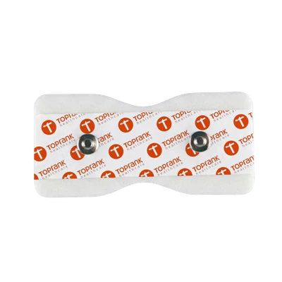
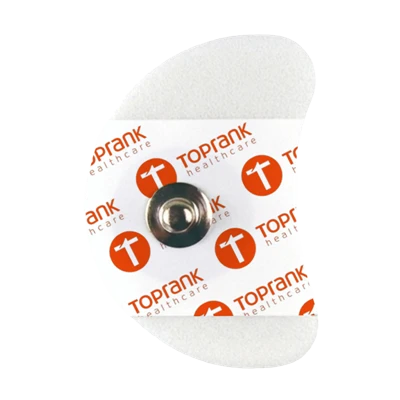
The sensing core utilizes an offset (eccentric) geometry. A precision brass snap stud (coated with nickel or silver) is staked into a high-density ABS plastic backing plate located 15 mm away from the active gel cavity. A flexible, screen-printed Ag/AgCl conductor trace bridges the offset snap to the central sensing well. The sensing well houses an ABS sensor eyelet coated with a 10 um thick, electrochemically deposited Ag/AgCl layer (Ag:AgCl ratio maintained at 85:15), delivering low half-cell potential variance and a baseline DC offset under 0.5 mV.
The active conductive medium consists of a high-chloride aqueous liquid gel (chloride concentration 3.0% to 4.5% by weight) held inside a circular disk of 60-PPI open-cell reticulated polyurethane foam. Liquid gel features low dynamic viscosity (<500 cP), allowing the electrolyte to wet the valleys of human skin micro-relief within 15 seconds, creating an ohmic conductive bridge. The polyurethane sponge acts as a fluid reservoir and mechanical baffle, preventing the gel from squeezing out under motion.
The central sponge well is sealed by a high-barrier circular PET film ring to prevent lateral gel migration into the surrounding adhesive zone during storage. Finished electrodes are mounted in arrays of 5 or 10 units on siliconized polyester (PET) release liners and sealed inside airtight PET/AL/PE multi-layer barrier pouches. Automated inline cameras verify snap staking pull force (>60N axial), sponge positioning, and liquid gel dosage (+/- 0.05 g). The standard contract manufacturing MOQ is 300,000 pieces.
Key Features
-
Offset Snap Mechanical Decoupling (Isolating Leadwire Dynamic Drag from the Electrochemical Double Layer)
During treadmill stress testing at speeds above 8.0 km/h, the patient's arm swing and torso movement whip the heavy monitor leadwires cyclically. In standard centered-snap electrodes, the snap stud sits directly atop the conductive gel column. Every jerk of the cable imparts lateral shearing and vertical peeling moments directly onto the skin-gel interface. This mechanical disturbance alters the thickness and charge density of the Helmholtz electrical double layer formed between the chloride ions and epidermal electrolytes. The resulting step-voltage artifact (V = Q / C) swings into the ECG preamplifier with amplitudes of 0.2 mV to 1.5 mV in the 0.1 Hz to 2.0 Hz frequency band-directly overlapping the ST segment and T wave. Our design locates the brass snap stud 15 mm away from the sensing well on a dedicated high-density ABS support platform. When the lead cable pulls, the kinetic stress dissipates across the mechanical foam backing. The central Ag/AgCl sensor core and liquid gel reservoir remain undisturbed against the skin, preserving the electrochemical double layer and delivering a stable, artifact-free ST segment during peak physical exertion.
-
Reticulated Foam Matrix with Aqueous Liquid Gel (Sub-15-Second Stratum Corneum Wet-Out and Sub-100 Ohm Baseline Impedance)
Solid hydrogels rely on slow polymer hydration: water molecules bound within an acrylic network must slowly diffuse through sebum and dry keratin scales, taking 5 to 10 minutes to bring contact impedance below 2,000 Ohm. In busy cardiac stress test labs, clinicians apply electrodes and start the treadmill protocol immediately, resulting in noisy, wandering baselines during the crucial early stages. Our system utilizes an aqueous liquid electrolyte gel formulated with a low dynamic viscosity (<500 cP) and high ionic strength, held in a 60-PPI reticulated open-cell polyurethane foam disk. When pressed against the skin, capillary pressure forces the liquid gel out of the open sponge pores into the microscopic epidermal crevices. The high-mobility saline solution hydrates dry keratinocytes within 15 seconds, collapsing interface resistance. Small-signal AC impedance at 10 Hz drops below 100 Ohm (compared to 800 to 2,000 Ohm for solid gels). The ECG workstation acquires clean, high-amplitude QRS complexes and crisp P-waves from the first seconds of treadmill movement without requiring abrasive skin prep.
-
Hydrophobic Crosslinked Acrylic Boundary Barrier (Resisting Profuse Diaphoretic Delamination Under High-Velocity Treadmill Shear)
Profuse sweating presents a severe mechanical hazard in cardiac ergometry: sweat fluids pool beneath the adhesive border, weakening standard pressure-sensitive adhesives via hydro-plasticization until the electrode detaches. Our platform utilizes a synthetic crosslinked pure acrylic adhesive compounded without hydrophilic tackifiers. The adhesive exhibits high hydrophobic water repellency: when perspiration emerges from eccrine sweat ducts around the electrode border, the hydrophobic barrier halts capillary wicking across the adhesive plane. The closed-cell crosslinked PE foam backing (1.0 mm thickness, density 70 kg/m3) functions as a fluid gasket, maintaining a seal around the central sponge cavity while the outer perimeter anchors into the dry perimeter skin with a dynamic peel force exceeding 6.5N / 25mm. In continuous treadmill tests running through Bruce Protocol Stages 4 and 5 (heart rates >160 bpm, profuse diaphoresis, ambient 25C), the electrode retains over 85% of its initial adhesive retention force, eliminating lead detachment and mid-test aborted procedures.
Applications
Cardiology Outpatient Exercise Tolerance Testing
Routine 12-lead diagnostic telemetry electrodes for Bruce, Modified Bruce, and Balke treadmill ergometry protocols, detecting exercise-induced myocardial ischemia and coronary artery disease (CAD) without motion-induced ST-segment false alarms.
Emergency Department & Chest Pain Units (Diaphoretic ACS Triage)
Rapid-application diagnostic monitoring for patients arriving with acute myocardial infarction (STEMI/NSTEMI), cardiogenic shock, or severe vasovagal diaphoresis, establishing immediate electrical contact through cool, clammy sweat without skin prep.
Sports Medicine & Elite Athletic Physiology Laboratories
Multi-stage VO2-max cardiopulmonary exercise testing (CPET) on elite cyclists and runners, withstanding intense physical vibration, high skin friction, and sweat rates exceeding 2.0 L/h through 60-minute peak exhaustion runs.
OEM & Private Label
-
Footprint Geometries, Pouch Formats & Turnkey Brand Customization : We offer custom tooling for teardrop, oval, and dual-snap stress testing geometries mounted on siliconized PET release liners. The packaging features an impermeable PET/AL/PE foil pouch with multi-layer barrier seals to prevent liquid gel dry-out. Pouch configurations include 3, 5, 10, or 30 electrodes per pack, matching 10-lead or 12-lead stress cable harnesses. Foam backings can be custom surface-printed with private-label brand logos, lead placement identifiers, or barcode/UDI traceability markers.
-
Two-Center Supply Chain Mechanics : High-precision Ag/AgCl electrochemical eyelet deposition, automated high-speed PE foam die-cutting, offset brass snap staking, and liquid gel precision micro-metered dispensing execute natively inside our primary China facility. Secondary foil pouch hermetic sealing, dynamic impedance batch validation, visual defect camera inspection, cleanroom sterile packaging, and export container palletization route through our Vietnam hub, shielding international B2B buyers from regional medical consumable tariffs and Section 301 duties.
Certifications
-
ANSI/AAMI EC12 & IEC 60601-2-27 Electrical Verification : Master production lots undergo testing in accredited independent testing laboratories confirming strict compliance with ANSI/AAMI EC12 (Disposable ECG electrodes) and IEC 60601-2-27. Test protocols confirm initial DC offset voltage remains below 1.0 mV (typical <0.5 mV), small-signal AC impedance remains below 100 Ohm at 10 Hz, and post-defibrillation overload recovery settles below 20 mV within 5.0 seconds.
-
ISO 10993 Dermal Biocompatibility Rigor : Direct skin-contact materials, including liquid gel sponges and acrylic adhesives, undergo batch validation against ISO 10993-5 (In Vitro Cytotoxicity Grade 0), ISO 10993-10 (Skin Sensitization: 0% allergic response), and ISO 10993-23 (Primary Dermal Irritation Index = 0.0). The complete assembly is 100% natural rubber latex-free, phthalate-free, and solvent-free.
-
MDSAP & ISO 13485 Manufacturing System Controls : High-speed converting lines, automated eyelet staking decks, and barrier sealing suites operate strictly within facilities certified to ISO 13485:2016 and MDSAP regulatory frameworks (covering US FDA 21 CFR 820, Health Canada, TGA Australia). Unbroken electronic Device History Records (DHR) log all critical manufacturing parameters, including raw foam peel values, Ag/AgCl coating thickness, liquid gel dispense volumes, and final pouch seal integrity.
Technical FAQ
Q: Why does aqueous liquid gel (wet gel) outperform solid hydrogel in dynamic treadmill stress testing, and how does the reticulated sponge reservoir prevent gel weeping under high impact?
A: Solid hydrogels are chemically cross-linked viscoelastic solids where water molecules are bound within polymer chains. Because bound water exhibits low thermodynamic mobility, a solid hydrogel requires 5 to 10 minutes of contact pressure to soften the lipophilic stratum corneum. During this hydration lag, interface resistance remains high (Rct > 1,500Ω). If the treadmill test starts immediately, this high baseline resistance causes severe baseline wandering and noise artifact. In contrast, our liquid gel is an aqueous solution featuring high concentrations of dissociated sodium and chloride ions unconstrained by a polymer backbone. It achieves instantaneous stratum corneum penetration, dropping the charge-transfer resistance below 100 Ohm within 15 seconds of application. The mechanical challenge of liquid gel is weeping (fluid leakage under pressure): when a patient runs, thoracic expansion and clothing friction apply cyclical compressive shocks that can squeeze liquid out of standard foam wells, shorting adjacent leads or causing adhesive failure. We eliminate weeping by holding the gel inside a 60-PPI open-cell reticulated polyurethane foam sponge. The microscopic dodecahedral skeletal cells create strong capillary capillary forces (Pc = 2γcosθ/r). These capillary forces hold the liquid gel firmly within the sponge matrix, preventing fluid leakage even under 15 kPa dynamic compressive shocks while allowing the fluid to maintain a wet conductive contact plane against the skin.
Q: How does the offset snap geometry eliminate ST-segment elevation/depression artifacts caused by leadwire cable drag during peak exertion (Stage 4+ Bruce protocol)?
A: During Stage 4 of the Bruce protocol (speed: 6.8 km/h, grade: 16%), patient acceleration and vertical oscillation reach peak values. Heavy trunk cables (typically 10-lead shielded harnesses weighing 200 to 300 grams) bounce vigorously. In standard centered-snap electrodes, the snap eyelet is located directly above the Ag/AgCl sensor and conductive gel column. The swinging cable acts as a mechanical lever: axial pull forces apply cyclic peeling moments (M = F × d) directly to the conductive core. This micro-mechanical displacement deforms the Helmholtz electrical double layer formed at the Ag/AgCl-to-gel and gel-to-epidermis boundaries. Because the double layer acts as a charged capacitor with a fixed interfacial charge (Q), changing the contact geometry alters the capacitance (C), generating an artifactual voltage shift:Δ V = QΔ CThis transient potential shift occurs at 1.0 to 3.0 Hz (the running stride frequency), exactly mirroring the frequency spectrum of cardiac repolarization (ST segment and T wave). Clinicians misinterpret these cable-drag voltage drops as true ischemic ST-segment depression. Our offset snap geometry moves the metal snap stud 15 mm away from the conductive core onto an independent PE foam extension backed by a rigid ABS plate. When the lead cable pulls, the tensile and peeling stresses are completely absorbed and dissipated by the outer foam backing and adhesive perimeter. The active Ag/AgCl eyelet and sponge well experience zero mechanical shear, holding the capacitance (C) of the Helmholtz double layer constant. The baseline remains flat, and ST segments reflect authentic myocardial perfusion without motion-induced diagnostic errors.
Cardiovascular diagnostic equipment manufacturers, exercise stress testing laboratory directors, and emergency medical consumable distributors can request accredited ANSI/AAMI EC12 electrical compliance dossiers, dynamic treadmill motion-artifact oscillograms, and unbranded evaluation sample pouches (teardrop offset, 5 or 10 pcs/pack).
👉 [Request Stress Test & Diaphoretic Electrode Evaluation Packs]
Hot Tags: stress test / diaphoretic ecg electrodes, China stress test / diaphoretic ecg electrodes manufacturers, suppliers, factory, Ecg Electrode, Ecg Electrode Stickers, electrode stickers
Specifications
| Analytical Parameter | Engineering Baseline | Testing Standard |
| Regulatory Mandate | ANSI/AAMI EC12:2000/(R)2020, IEC 60601-2-27 | Disposable ECG electrodes |
| Electrode Footprint | Teardrop Offset: 40 mm x 50 mm or 45 mm x 55 mm | Ergonomic profile |
| Terminal Interface | Off-Center Solid Brass Stud with Nickel/Silver Plate | Offset mechanical decoupling |
| Sensor Element | High-Purity Ag/AgCl Coated on ABS Substrate | Reversible redox pair |
| Conductive Medium | Aqueous Chloride Liquid Gel in Reticulated PU Foam | Fast-wetting wet gel |
| AC Small-Signal Impedance | < 100 Ohm at 10 Hz (EC12 limit: < 3,000 Ohm) | Four-wire test setup |
| DC Offset Voltage | < 1.0 mV (Typical < 0.5 mV, EC12 limit: < 100 mV) | Unbalanced half-cell |
| Baseline Drift Rate | < 0.1 mV/s over 30 Minutes of Dynamic Exertion | In-vitro motion fixture |
| Defibrillation Recovery | Residual offset < 20 mV after 5.0 seconds | Post-shock pacing |
| Dynamic Peel Adhesion | > 6.5N / 25 mm on Polished Stainless Steel | ASTM D3330 |
| Moisture Resistance | 60 Minutes Continuous at 38C and 90% RH (Sweat Test) | Artificial sweat immersion |
| Substrate Backing | 1.0 mm Closed-Cell Crosslinked PE Foam (70 kg/m3) | Fluid-impermeable barrier |
| Packaging & Pouching | Sealed Multi-Layer PET/AL/PE Foil Pouch (5 or 10 pcs) | 24-Month shelf life |