

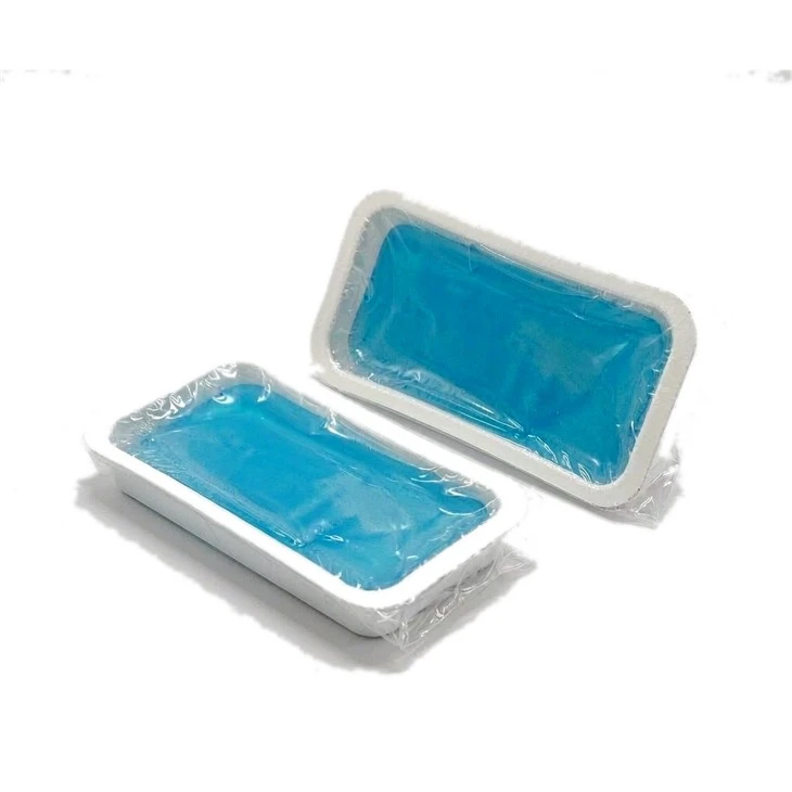
OEM AutoGel Pads | FDA 510(k) Cleared Ultrasound Gel Pads | TOP-RANK
B2B contract manufacturing of AutoGel Pads (Ultrasound Standoff Gel Pads) cleared under US FDA 510(k) K260575. Delivers acoustic velocity 1540 m/s, impedance 1.54 MRayls, attenuation <0.08 dB/cm-MHz, mess-free solid standoff, and dual China-Vietnam export routing.
Product Overview
AutoGel Pads function as pre-formed, solid-phase acoustic standoff hydrogel pads designed for high-resolution diagnostic ultrasound examinations. Formulated as a mess-free alternative to bulk liquid couplants, these elastomeric gel slabs couple diagnostic ultrasound transducers to irregular, bony, or painful patient anatomy without requiring bottles of runny gel. The pad displaces the transducer's near-field reverberation dead-zone (typically the first 0 mm to 10 mm beneath the probe lens) out into the hydrogel standoff mass. This shifts superficial biological targets-such as thyroid nodules, superficial breast lesions, dermal melanomas, foreign bodies, infant cranial windows, and musculoskeletal tendons-directly into the probe's optimal focal zone. Validating its acoustic fidelity, physical safety, and manufacturing quality controls, this product platform has officially received US FDA 510(k) clearance under submission number K260575 (clearance granted; public FDA database index synchronization pending).
Performing superficial and musculoskeletal (MSK) scans with conventional liquid ultrasound gels or low-grade gelatin pads introduces three persistent diagnostic and mechanical failures:
- Near-Field Reverberation Ghosting and Artifact Blindness: Standard linear ultrasound probes (operating between 7.5 MHz and 22 MHz) exhibit an inherent physical acoustic dead-zone directly adjacent to the transducer acoustic lens. In this zone, intense reflection echoes and transducer ringing obscure biological structures located in the first 1 to 5 mm of the dermis. When sonographers apply liquid gel directly to the skin, superficial pathologies (like digital flexor tendon tears or epidermal cysts) remain hidden inside this near-field noise band, leading to missed diagnoses.
- Liquid Gel Slumping and Air Crevicing on Bony Contours: Examining irregular, angular surfaces (such as the acromioclavicular joint, knuckles, malleolus, open surgical incisions, or the Achilles tendon) requires flat transducer contact. Liquid ultrasound gel has low yield stress; it runs off sloping anatomy, leaving dry patches. Tilted probes trap air pockets in skin crevices, causing total acoustic reflection (acoustic shadowing) that obscures deep tissue architecture and forces technicians to exert excessive probe pressure, causing acute pain to inflamed joints.
- Internal Acoustic Inhomogeneity and Probe Reverberation: Low-cost molded gel blocks manufactured from agar, silicone, or poorly crosslinked polymers have uneven internal density distributions and entrapped micro-voids. When high-frequency sound waves traverse these flawed media, acoustic impedance mismatches generate internal reverberation artifacts (multiple reflection banding). Furthermore, high polymer viscous damping attenuates 15+ MHz ultrasound frequencies, muddying fine anatomical margins and compromising Doppler flow signals.
The AutoGel Pad platform resolves these acoustic and clinical limitations through specialized polymer engineering: a crosslinked polyacrylamide-co-polyacrylate solid hydrogel matrix synthesized with an exact acoustic velocity of 1540 m/s (+/- 10 m/s) and an acoustic impedance of 1.54 MRayls at 25C to 37C, matching human soft tissue; a clean vacuum-cast manufacturing process that achieves an internal micro-bubble count of zero, limiting acoustic attenuation below 0.08 dB/cm-MHz across 5 MHz to 22 MHz probes; and a tear-resistant, high-cohesion slab (thickness: 10 mm to 20 mm) that flexes around sharp bone contours without liquefying or leaving messy residue on probes or skin. Cleared under FDA 510(k) K260575, we contract-manufacture private-label AutoGel Pads in sterile and non-sterile blister packs for medical imaging OEMs, hospital procurement consortiums, and ultrasound consumable distributors globally.
Technical & Engineering Description
The industrial synthesis and automated casting of AutoGel Pads execute inside ISO 13485-certified Class 8 cleanrooms across dedicated fluid-degassing reactors and automated thermoformed blister-dispensing lines operating under US FDA 21 CFR 820 design controls. Production proceeds across five coordinated engineering stages:
- Stage 1: Multi-Pass Water Deionization & Acoustic Monomer Compounding: Pharmaceutical raw water is purified through dual-pass reverse osmosis (RO) and continuous electrodeionization (EDI), suppressing conductivity below 0.1 uS/cm. The polished water charges into jacketed 316L stainless steel reaction vessels. Analytical-grade acrylamide, acrylic acid, biocompatible covalent crosslinkers (methylenebisacrylamide), and organic humectants are added in exact stoichiometric ratios. The formulation compounds an inorganic salt buffer that balances sound speed to 1540 m/s and acoustic impedance to 1.54 MRayls, harmonizing the acoustic properties of the fluid with human tissue.
- Stage 2: High-Vacuum Centrifugal Deaeration & Bubble Scrubbing: Microscopic air bubbles act as severe acoustic reflection and scattering barriers that degrade ultrasound waves. The compounded monomer solution transfers into an industrial vacuum degassing chamber drawn down to an absolute negative pressure of -0.098 MPa under continuous planetary centrifugal rotation (1,800 RPM). Centrifugal force and high vacuum pull micro-air bubbles and dissolved atmospheric gases out of the high-viscosity liquid, continuing until dissolved oxygen drops below 0.2 ppm and internal micro-bubble counts drop to zero under automated laser obscuration particle counters.
- Stage 3: Automated In-Blister Dispensing & Inert Thermal Polymerization: The deaerated monomer solution routes through sanitary positive-displacement ceramic metering pumps. The liquid dispenses directly into pre-formed, medical-grade PETG rigid blister trays at calibrated volumetric doses corresponding to finished pad thicknesses of 10.0 mm, 15.0 mm, or 20.0 mm (+/- 0.5 mm). The filled blister trays enter an enclosed nitrogen-purged thermal curing tunnel operating below 40 ppm oxygen. Controlled thermal profiles (65C to 80C) trigger a uniform free-radical copolymerization across 45 minutes, transforming the liquid monomer into a tough, solid-phase hydrogel slab molded directly to the shape of the blister cavity.
- Stage 4: Optical Laser Inspection & Automated Hydrostatic Acoustic Profiling: De-molded and in-tray gel slabs pass through an automated quality-assurance deck. High-resolution line-scan cameras verify edge contour concentricity, thickness uniformity, and absolute optical transparency (confirming absence of micro-voids or turbidity). Every master lot undergoes automated acoustic parameter testing using calibrated 10 MHz ultrasound transducers: measuring through-transmission transit time to confirm sound speed at 1540 m/s (+/- 10 m/s), measuring acoustic impedance, and recording attenuation coefficients across a 5 MHz to 20 MHz spectrum.
- Stage 5: Tyvek/Foil Barrier Sealing & Terminal Sterilization Routing: The blister trays receive breathable medical-grade Tyvek lids (for radiation sterilization) or multi-layer aluminum barrier foil peel-lids (for standard shelf stability). Ultrasonic and thermal platen sealers apply 0.45 MPa pressure, creating hermetic, leak-proof perimeter seals. For intra-operative and biopsy-guided applications, sealed trays route through gamma radiation sterilization (validated to ISO 11137 achieving a Sterility Assurance Level of 10^-6). Finished trays box into clinical master cartons with serialized GS1/UDI barcodes. The baseline private-label contract manufacturing MOQ is 10,000 units.
Key Features
-
1540 m/s Human Tissue Acoustic Matching (Shifting Transducer Near-Field Reverberation Out of the Diagnostic Window)
The defining diagnostic limitation of high-frequency ultrasound in superficial scanning is transducer near-field reverberation noise:
1. The Physics of the Acoustic Dead-Zone: Medical ultrasound probes generate acoustic wave packets via piezoelectric crystals positioned immediately behind an acoustic lens. During pulse emission, the piezoelectric elements ring violently. This high-amplitude mechanical ringing, coupled with sound wave bounce between the lens and skin, creates a continuous band of chaotic reverberation noise that saturates the receiving amplifiers for the first 0 mm to 10 mm of acoustic path length (the "acoustic dead-zone").
2. Superficial Pathology Masking: When diagnosing conditions located just beneath the skin-such as a 2 mm superficial melanoma, digital flexor tendon tears, infant skull cranial sutures, or early-stage thyroid capsule microcalcifications-the lesion sits directly inside this noisy dead-zone. In conventional scans using liquid gel, the pathology is masked by bright, stripe-like reverberation artifacts.
3. Acoustic Distortion in Generic Standoffs: Cheap silicone or agar standoff blocks have sound speeds differing significantly from tissue (e.g., silicone conducts sound at ~1000 m/s, whereas tissue is ~1540 m/s). This speed mismatch introduces refraction distortion, incorrect depth calculations, and severe ghost reflections at the boundary. Our AutoGel Pad platform resolves near-field blindness through true acoustic impedance and speed matching:
4. Tissue-Identical Sound Speed: Compounded with an organic electrolyte-water grid, the AutoGel matrix transmits longitudinal acoustic waves at an exact velocity of 1540 m/s (+/- 10 m/s) with a matched acoustic impedance of 1.54 MRayls.
5. Near-Field Relocation: By placing a 10 mm or 20 mm AutoGel Pad between the probe and patient, the entire acoustic dead-zone is displaced outward into the sterile mass of the gel pad.
6. Pristine Focal-Zone Imaging: The true patient epidermis and superficial subcutaneous tissues now enter the sound beam at a depth of 10 mm to 20 mm, falling directly into the probe's electronic focal point. Reverberation noise is eliminated, rendering skin layers, tendon bundles, and sub-millimeter foreign bodies with high diagnostic contrast and zero depth distortion.
-
Conformal Bony-Contour Adaptability (Eliminating Acoustic Shadowing from Trapped Air on Knuckles, Ankles, and Wounds)
A frequent clinical challenge in musculoskeletal (MSK) and emergency ultrasound is acoustic wave obstruction across angular, non-planar anatomical contours:
1. The Air Reflection Physics: Air has an acoustic impedance of only 0.0004 MRayls, compared to 1.54 MRayls for human tissue. The reflection coefficient at an air-tissue boundary is nearly 100%. When an ultrasound wave encounters even a microscopic pocket of trapped air, the entire acoustic beam reflects back, casting a dark acoustic shadow that blinds the monitor to all tissue structures beneath the air void.
2. Transducer Bridging on Bony Landmarks: When sonographers evaluate curved, bony anatomy-such as metacarpophalangeal joints, malleoli, clavicles, or open post-surgical suture ridges-the flat acoustic lens of the probe bridges across bony ridges. Liquid gel quickly runs down the sides of the joint, leaving air pockets beneath the probe center. The technician is forced to push the hard probe firmly against the skin, crushing sensitive structures and causing acute pain to patients suffering from acute fractures or rheumatoid arthritis. Our AutoGel Pad platform eliminates air-pocket shadowing through mechanical compliance and surface wet-out:
3. High-Elongation Interpenetrating Lattice: The synthetic hydrogel balances tensile strength (>0.8 MPa) with high elastomeric elongation (>300%), allowing the thick slab to deform and drape around angular anatomical structures (such as wrapping 90 degrees around an ankle or knuckle).
4. Zero-Pressure Air Displacement: Under light probe contact, the compliant gel flows into micro-valleys around skin creases, bony humps, and suture lines, displacing air pockets without requiring downward compression.
5. Pain-Free "Floating Probe" Scanning: The sonographer can float the probe lightly across the top surface of the gel pad without applying direct pressure to the patient's inflamed skin, providing pain-free diagnostic imaging across tender fractures, burns, and post-operative surgical incisions.
-
Zero-Residue Mess-Free Clinical Workflow (Eliminating Probe Disinfection Slime and Patient Post-Scan Clean-Up Overhead)
In high-throughput ultrasound departments and emergency triage suites, time spent cleaning equipment directly impacts patient turnover:
1. The Mess of Liquid Couplants: Applying bottles of liquid ultrasound gel coats the patient's skin and hair in a cold, sticky fluid. Following the scan, technicians consume piles of paper towels attempting to wipe the gel off. Residue frequently remains stuck in hair follicles or smears onto patient clothing, creating patient discomfort.
2. Transducer Contamination & Degradation: Liquid gel seeps into microscopic seams around the acoustic lens, cable strain-relief joints, and freeze-frame button crevices of the transducer. Over time, volatile solvents and salts in the gel degrade the silicone adhesive sealing the acoustic lens. Fluid ingress initiates internal electrical corrosion of the piezoelectric array, resulting in costly probe refurbishment (often 5,000 to 10,000 per transducer) and risk of cross-contamination between patients. We eliminate coupling clean-up overhead through clean solid-phase cohesion:
3. High-Cohesion Solid Polymer Network: AutoGel Pads are manufactured as unified, cohesive solid hydrogel slabs that contain zero loose, unbonded liquid phase. The material possesses high internal cohesive energy that exceeds its surface peel adhesion.
4. Zero-Residue Lift-Off: Following the ultrasound examination, the technician peels the AutoGel Pad off the patient's skin in a single, smooth pull. The pad releases cleanly, leaving the patient's skin, hair, and clothing dry with zero slimy film or sticky residue (wipe residue = 0.0 mg).
Pristine Transducer Lens: The ultrasound probe remains dry throughout the procedure. There is zero liquid to seep into probe seams, protecting the acoustic lens seal from chemical degradation and allowing instant wipe-down disinfection with hospital surface wipes between patients, cutting post-scan room turnover time by more than 60%.
Applications
Superficial Small-Parts Diagnostic Imaging (Thyroid, Breast & Pediatric Cranial Windows)
Sourced in high container volumes by hospital radiology departments, outpatient imaging centers, and maternal-fetal specialty suites; utilizing standard 10 mm and 15 mm rectangular AutoGel Pads beneath 12 MHz to 18 MHz linear array probes; the pad shifts the transducer acoustic dead-zone completely outside the patient, allowing radiologists to assess thyroid capsule micro-invasions, non-palpable breast intraductal papillomas, and neonatal ventricular morphology with sharp border resolution.
Musculoskeletal (MSK) Sports Medicine & Rheumatology Joint Profiling
Deployed across orthopedic sports medicine clinics, collegiate athletic training rooms, and rheumatology specialty practices evaluating soft-tissue injuries; the high-flexibility AutoGel matrix contours over metacarpophalangeal joints, lateral malleoli, and the Achilles tendon, eliminating air-pocket shadowing while allowing pain-free "floating-probe" scanning without putting mechanical pressure on acutely sprained ligaments or inflamed synovial joints.
Emergency Department Trauma, Burn Staging & Sterile Biopsy Guidance
Stocked across emergency department trauma suites, burn intensive care units (BICU), and interventional radiology operating rooms; gamma-sterilized AutoGel Pads (ISO 11137 verified) place an impermeable acoustic barrier directly over open lacerations, fresh surgical incisions, and partial-thickness burn beds, enabling emergency physicians to guide needle insertions, locate radiolucent foreign bodies, and stage tissue viability without introducing infection or stinging injured skin.
OEM & Private Label
-
Pad Geometries, Sterile/Non-Sterile Assortment & Custom Packaging : We provide comprehensive contract manufacturing and formulation packaging options for medical imaging brands and clinical consumable distributors:
• Footprint Geometry Matrix: Standard Square in 100x100 mm and 150x150 mm (wide-field linear probe coverage); Classic Circular in Dia 100 mm (for rotating vascular probes); Anatomical Contoured Strips (custom curved shapes for flexor tendons and orbital scanning via dedicated tooling).
• Thickness Profiling Matrix: Standard 10.0 mm (universal near-field shift for 10-15 MHz probes); Extended 15.0 mm (for ultra-high-frequency 18-22 MHz dermatology probes); and Heavy 20.0 mm (for deep tendon clearance and foreign-body extraction protocols).
• Aseptic & Clinical Packaging Options: Available in non-sterile clinical economy multipacks (sealed inside rigid thermoformed PETG blister trays with reclosable foil lids); or terminal gamma-sterilized individual peel-pouches (ISO 11137 verified with medical Tyvek lidding for direct delivery into sterile surgical fields).
• Private-Label Packaging: Multi-color offset outer carton printing, custom brand logos directly hot-stamped on blister foil lids, clinical IFU brochures with anatomical placement diagrams, and serialized GS1/UDI barcodes.
-
Two-Center Supply Chain Mechanics : High-purity water polishing, proprietary AutoGel monomer chemical synthesis, high-vacuum centrifugal degassing reactors, and automated cleanroom thermoforming cast lines execute natively inside our primary China facility (ISO 13485:2016 and FDA registered under 21 CFR 820). High-speed automated blister sealing, machine vision quality inspection, secondary sterile cartoning, and palletized ocean container export route through our Vietnam manufacturing hub, shielding international B2B buyers from Section 301 tariffs and regional medical consumable import barriers.
Certifications
-
US FDA 510(k) Premarket Clearance (K260575) : The AutoGel Pad (Ultrasound Standoff Gel Pad) platform has satisfied premarket notification requirements under Section 510(k) of the Federal Food, Drug, and Cosmetic Act, obtaining official clearance under K260575 (classified under 21 CFR 892.1570, Diagnostic Ultrasound Transducer Acoustic Standoff Couplant). The regulatory technical dossier confirms substantial equivalence in acoustic velocity matching, ultrasonic attenuation, bubble-free transmission, and biocompatibility against established predicate devices.
-
ISO 10993 Comprehensive Dermal Biocompatibility : Master production lots undergo testing in accredited independent GLP testing laboratories confirming strict compliance with ISO 10993-5 (In Vitro Cytotoxicity: Grade 0 non-cytotoxic), ISO 10993-10 (Skin Sensitization: 0% allergic response in guinea pig maximization assays), and ISO 10993-23 (Primary Dermal Irritation Index = 0.0). The hydrogel matrix is certified 100% natural rubber latex-free, animal-gelatin-free, phthalate-free, and safe for direct application over damaged or sensitive skin.
-
ISO 11137 Radiation Sterilization Validation (Sterile Product Lines) : For sterile product lines, packaging lots undergo validation under ISO 11137-1 and ISO 11137-2 (Sterilization of health care products - Radiation). Dose-setting audits confirm that the validated gamma sterilization dose achieves an unbroken Sterility Assurance Level (SAL) of 10^-6 with zero degradation of acoustic sound velocity or mechanical elasticity.
-
MDSAP & ISO 13485 Manufacturing System Controls : Monomer synthesis reactors, degassing chambers, and blister heat-sealing bays operate strictly under certified ISO 13485:2016 and MDSAP regulatory frameworks (covering US FDA 21 CFR 820, Health Canada, TGA Australia). Unbroken electronic Device History Records (DHR) capture bulk deaeration vacuum levels, ultrasound sound-velocity verification logs, acoustic attenuation charts, and blister seal burst-pressure data across every manufactured production lot.
Technical FAQ
Q: How does the FDA 510(k) K260575 cleared AutoGel Pad eliminate near-field reverberation noise on 15 MHz linear probes compared to standard liquid gels?
A: The elimination of near-field reverberation artifacts is governed by physical acoustic path displacement and matched sound velocity impedance: The Physics of Near-Field Reverberation Noise: Diagnostic linear ultrasound probes emit high-frequency acoustic wave packets from an array of piezoelectric crystals positioned behind a protective acoustic lens. When the piezoelectric elements fire: 1. The crystals experience mechanical resonance ringing, generating high-amplitude acoustic reverberations directly adjacent to the probe surface. 2. Sound waves bounce back and forth between the acoustic lens and the patient's skin, saturating the receiving electronics for the first 0 mm to 10 mm of depth (the "acoustic dead-zone"). 3. When an ultrasound technician attempts to scan structures located directly beneath the epidermis (such as an Achilles tendon tear, superficial breast cyst, or digital nerve), these micro-structures sit inside this noisy dead-zone. In conventional imaging using thin liquid gels, the pathology is obscured by bright, horizontal reverberation artifact lines. Our AutoGel Standoff Displacement Engineering: The AutoGel Pad (cleared under FDA 510(k) K260575) eliminates this artifact through acoustic path length relocation: 1. Relocating the Dead-Zone Outside the Body: The 10 mm to 20 mm thick solid AutoGel Pad acts as a physical acoustic spacer. The entire chaotic reverberation zone occurs inside the sterile, uniform mass of the gel pad itself, before the sound beam ever enters the patient's body. 2. Matching Acoustic Velocity (1540 m/s): Because the AutoGel matrix transmits sound at an exact 1540 m/s with an acoustic impedance of 1.54 MRayls, there is zero impedance mismatch at the pad-skin boundary. Sound waves transition smoothly from the gel pad into human tissue without generating secondary interface echoes. 3. Positioning Superficial Pathology into the True Focal Zone: The true anatomical epidermis and subcutaneous tissues enter the acoustic beam at a depth of 10 mm to 20 mm, falling directly into the probe's electronic focal point. The reverberation artifact disappears, revealing superficial lesions with clear acoustic margins and high diagnostic contrast.
Q: Why does high-frequency ultrasound lose signal strength (acoustic attenuation) through cheap standoff pads, and how does AutoGel maintain Doppler blood flow sensitivity?
A: Acoustic energy loss through standoff pads is governed by polymer viscous damping and ultrasonic scattering from internal micro-inhomogeneities: The Attenuation Breakdown of Low-Cost Standoffs: Low-cost acoustic standoffs molded from consumer-grade silicones, agars, or rubber hydrogels exhibit severe acoustic attenuation when subjected to high-frequency diagnostic sound waves (10 MHz to 20 MHz): 1. Viscous Damping: Synthetic polymers with loose, irregular crosslinking have high internal viscoelastic damping. As high-frequency sound waves pass through the material, acoustic energy is converted into heat via internal molecular friction. In basic silicone pads, attenuation reaches 1.5 to 3.0 dB/cm-MHz. 2. Micro-Bubble Scattering: Open-mold cast pads trap microscopic air bubbles. These micro-voids act as acoustic scatterers, reflecting and refracting sound waves in random directions. 3. Loss of High-Frequency Signal & Doppler Clamping: Because high-frequency ultrasound attenuates exponentially with frequency, a 15 MHz pulse traveling through a 15 mm low-cost pad loses over 70% of its acoustic power before reaching the patient. The return echoes from tiny red blood cells are too weak to register, blinding color Doppler flow imaging across superficial digital vessels and thyroid micro-vascularity. Our Low-Attenuation AutoGel Engineering: AutoGel Pads maintain high acoustic energy transmission through closed-loop molecular and degassing controls: 1. Vacuum Centrifugal Deaeration: Liquid monomer precursors undergo high-vacuum centrifugal degassing at -0.098 MPa, reducing the internal micro-bubble count to zero. Ultrasound waves propagate through the medium without Rayleigh scattering. 2. Low-Damping Interpenetrating Network: The polyacrylamide-co-polyacrylate hydrogel is synthesized with a dense, highly elastic covalent network plasticized with pure deionized water, minimizing viscoelastic loss. 3. Ultra-Low Attenuation (<0.08 dB/cm-MHz): Across the full 5 MHz to 22 MHz clinical diagnostic spectrum, the attenuation coefficient remains strictly below 0.08 dB/cm-MHz. Sound waves traverse the 10 mm to 20 mm AutoGel Pad with over 92% acoustic power retention. Weak, microvolt-level Doppler shift signals from superficial digital arteries and thyroid nodules penetrate back to the probe, delivering sensitive color flow mapping and spectral Doppler waveforms without attenuation drop-out.
Medical ultrasound transducer manufacturers, radiology consumable procurement directors, and hospital tender managers can request official Certificates of Analysis (COA), US FDA 510(k) K260575 premarket clearance documentation, 5-20 MHz acoustic attenuation laboratory curves, ISO 10993 dermal biocompatibility dossiers, and unbranded evaluation sample packs (including 100x100 mm square formats in 10 mm and 15 mm thickness profiles).
👉 [Request FDA 510(k) K260575 Cleared AutoGel Pad Evaluation Packs]
Hot Tags: autogel pads, China autogel pads manufacturers, suppliers, factory
Specifications
| Analytical Parameter | Engineering Baseline | Testing Standard |
| Regulatory Clearance | US FDA 510(k) Cleared: K260575 (Acoustic Standoff Couplant) | 21 CFR 892.1570 |
| Footprint Formats | Standard Rectangular: 100x100 mm, 150x150 mm; Disk: Dia 100 mm | Clinical imaging norms |
| Standoff Thickness | 10.0 mm, 15.0 mm, and 20.0 mm (+/- 0.5 mm caliper control) | Caliper gauge |
| Speed of Sound | 1540 m/s (+/- 10 m/s at 25C to 37C) | ASTM E494 |
| Acoustic Impedance | 1.52 to 1.56 MRayls (Rayls x 10^6 kg/m2-s) | Reflection calculation |
| Acoustic Attenuation | < 0.08 dB/cm-MHz across 5.0 MHz to 22.0 MHz Frequency Band | Transmission insertion loss |
| Polymer Chemistry | Crosslinked Polyacrylamide-Co-Polyacrylate Hydrogel (IPN) | Synthetic network |
| Mechanical Integrity | Tensile Strength > 0.8 MPa; Elongation at Break > 300% | ASTM D412 |
| Surface Tack & Cleanliness | Balanced Conformal Self-Adhesion (Zero residue / Zero slip) | Probe wipe test |
| Optical Clarity | Crystal-Clear Transparent Matrix (Transmittance >92% at 632 nm) | UV-Vis Spectrophotometer |
| Biocompatibility Grade | Grade 0 Cytotoxicity; 0% Sensitization; Primary Irritation = 0.0 | ISO 10993 compliant |
| Sterility Options | Non-Sterile Clinic Packs or Gamma-Irradiated Sterile Packs | ISO 11137 (SAL 10^-6) |
| Primary Packaging | Rigid Thermoformed PETG Blister Trays with Tyvek/Foil Lids | 24 to 36-Month shelf life |
| Contract Manufacturing MOQ | 2,000 Pieces (Available in private-label clinical cartons) | Factory direct |


